Department head
Dr. Sabine Hombach-Klonisch
sabine.hombach-klonisch@umanitoba.ca
The University of Manitoba campuses and research spaces are located on original lands of Anishinaabeg, Ininiwak, Anisininewuk, Dakota Oyate, Dene and Inuit, and on the National Homeland of the Red River Métis. More
University of Manitoba
Winnipeg, Manitoba Canada, R3T 2N2
The histology laboratory provides services for researchers and students to process their tissues and cells for histopathological examination.We offer paraffin, cryo, and electron microscopy techniques including processing, embedding, sectioning, staining and imaging. We have an array of common and special histological stains available for specific projects. Selected immunofluorescent and immunohistochemical detection and special assays are also available upon request. Histomorphometry studies related to changes in histopathology of samples can be identified using new techniques in imaging and analysis.
We provide comprehensive support for your research projects, offering state-of-the-art techniques and personalized consultation.

Our team will meet with you to thoroughly discuss your project and provide guidance on the best methods to handle your samples. This collaborative approach ensures that we understand your specific research goals and can design a tailored plan to meet your needs.
After the consultation, you will receive a detailed estimate for approval, along with precise instructions on how to prepare your samples for optimal results. We will then schedule an appointment for sample drop-off.
To ensure the quality of your work, please note that samples must not be brought to the lab without a prior appointment.
For further information or to schedule a consultation, please don't hesitate to call us at 204-789-3508 or email histology_services@umanitoba.ca.
NOTE: While we offer the tools and expertise needed to prepare your samples for analysis, please note that the responsibility for analyzing and interpreting the resulting data lies with the principal investigator who provided the samples.
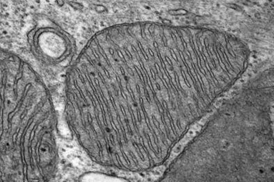
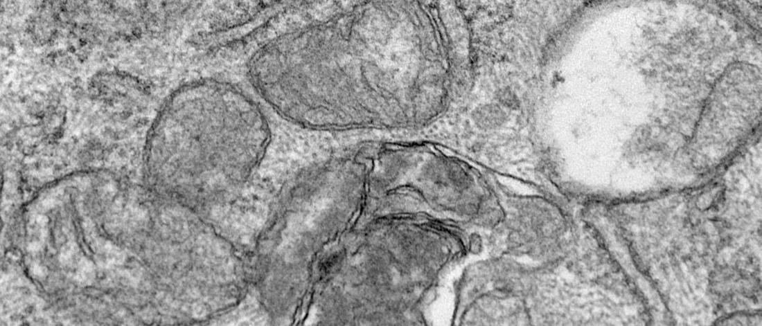
Our facility offers comprehensive electron microscopy to support researchers in processing biological tissues, cultured cells, bacteria, viruses, and nanoparticles for detailed ultrastructural analysis. We provide a full range of techniques, including tissue processing, embedding, sectioning, staining, and imaging, to meet the needs of your research projects.
Our consultations provide an opportunity for meaningful collaboration, where we discuss your project in detail, answer your questions, and design a tailored plan for processing your samples.
After your consultation, we’ll send an estimate for your approval. Once confirmed, we’ll guide you on how to prepare your samples for optimal results and schedule a convenient drop-off appointment. To ensure the best service we can only accept samples by appointment.
Our goal is to support your research every step of the way. From project planning to providing necessary fixatives and buffers for sample preparation, we take care of the details so you can focus on your work. Once we receive your samples, we handle everything else, delivering high-quality TEM images at your requested magnifications when your project is complete.
Please note that your samples should come with completed forms and a signed estimate.
To help you achieve the best possible results, please follow these guidelines for optimal tissue preparation:
Size:
Fixation:
Minimum fixation time:
To ensure thorough fixation, make sure your tissue samples are fully immersed and freely floating in a volume of fixative that is at least 10 times the volume of the tissue itself.
Our imaging facility is equipped with two state-of-the-art transmission electron microscopes (TEMs), providing researchers with the capability to observe subcellular changes. TEM techniques are invaluable for gaining deeper insights into how treatments, experimental conditions, or genetic modifications impact your samples, among many other factors.
We offer a variety of imaging and analysis tools to meet the diverse needs of your research, including:
Additionally, for a more quantitative analysis of experimental changes, we can incorporate machine learning techniques into the evaluation process, providing precise and reliable results.
The pricing provided below applies to UM and affiliated researchers. For industry-specific pricing, please contact us.
| Task | Description | Item | Cost |
|---|---|---|---|
| Buffers and fixative solutions | All necessary buffers and solutions for tissue or cell fixation, including a storage buffer. | Glutaraldehyde, Sorensen’s, phosphate buffer, sucrose | $30 (up to 50ml) |
| Labour (1 hour) | $50/hour | ||
| Tissue processing and embedding for EM | Post-fixation of tissue or cells (pre-grossed) through to embedding in resin. | Osmium tetroxide, alcohol dehydration, propylene oxide, EmBed 812 resin system | Included |
| 1-10 blocks | $250 | ||
| Labour (6-hour set) | $50/hour | ||
| Sectioning (diamond knife) | Thick sectioning with toluidine blue, thin sectioning (90-100nm) and grid placement (200 or 300 mesh copper). | Diamond knife, slides, toluidine blue, grids | Included |
| Labour | $50/hour | ||
| Grid staining | Staining with uranyl acetate, lead citrate, or uranyless. | Staining solutions | Included |
| Labour | $50/hour | ||
| Tem imaging (Phillips CM10) | Tem imaging by histology staff. | UM faculty and affiliates | $75/hour |
| External | $120/hour | ||
| USB | Saving images to USB for client | USB | $8 |
| Digital transfer | Digital image transfer via online platform | N/A | $0 |
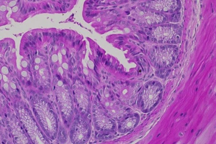
We provide comprehensive histology services, including standard paraffin embedding and frozen tissue preparation to support a wide range of biological research.
Our consultation process allows us to tailor our services to your specific research needs.
We utilize a wide range of advanced staining techniques, enabling us to provide detailed visual insights into protein abundance, distribution, and localization. These techniques, which include common histological stains as well as specialized methods, offer the precision required to highlight specific cellular and tissue components.
Through immunohistochemistry, immunofluorescence, and other advanced assays, we help you identify and analyze key biomarkers, ensuring that you gain a comprehensive understanding of your samples at both the structural and functional levels.
Whether you're exploring cellular dynamics, disease mechanisms, or developmental processes, our team is equipped to support your project with state-of-the-art staining methods and expert guidance.
We offer standard paraffin embedding preparation, a widely used and reliable technique for tissue preservation and sectioning.
This method involves embedding your samples in paraffin wax, allowing for long-term storage and optimal preservation of cellular architecture.
The process includes tissue fixation, dehydration, clearing, and embedding in molten paraffin, followed by sectioning into thin slices for histological examination. This approach is ideal for a wide range of tissue types and provides excellent preservation of morphological detail, making it suitable for routine histopathological analysis.
Additionally, our facility supports advanced staining techniques for further examination of tissue structure, protein localization, and other molecular characteristics, enabling a thorough investigation of your samples.
The pricing shown here is for UM and affiliated investigators. For industry pricing, please contact us.
| Task | Description | Item | Cost |
|---|---|---|---|
| Processing tissue for paraffin | Placing tissue that arrives in fixative (already grossed) into cassettes and labeling for the tissue processor | Fixative, formalin, dehydrating alcohol, xylene and paraffin | included in processing charge |
| 1-24 cassettes | $50 | ||
| Labour | $50/hour | ||
| Embedding tissue in paraffin | Taking the cassettes from the tissue processor and setting them in paraffin in the correct orientation, then filling the mold with paraffin to create the block | Paraffin from embedding machine | Included in tissue processing charge |
| Cutting paraffin sections | Sectioning the desired number for sections/slide from each block as requested | Superfrost plus | $1.80/slide |
| Knife blade | included | ||
| Labour | $50/hour | ||
| Staining paraffin sections | Staining and cover slipping slides as requested | Special stains | See staining page for special stains |
| H&E | $4/slide | ||
| Labour | $50/hour | ||
| IHC, IF and other assays | IHC or IF on the prepared slides with antibodies as well as other requested assays (kit) | Custom pricing for individual requests | $130/run plus all required supplies and chemicals |
| Labour | $50/hour | ||
| Imaging/analysis | Image the slides using brightfield or fluorescent microscope. (Taking pictures, performing analysis where required, counting, measuring, graphing) | UM | $50/hour self imaged $75/hour staff imaged |
| External/Industry | $75/hour self imaged $100/hour staff imaged | ||
| USB | Saving image to USB for client | USB | $8 |
| Digital transfer | Digital image transfer via online platform | N/A | $0 |
| Slide box | Slide storage/transport | Box | $15 |
For optimal results, proper preparation of your tissues is crucial. Adhering to these guidelines ensures the best possible outcomes for your frozen tissue samples:
By following these guidelines, you will ensure your tissues are optimally prepared for histological analysis and imaging, yielding the best results for your research.
The pricing shown here applies to University of Manitoba (UM) and affiliated researchers. For industry pricing or additional information, please contact us.
| Task | Description | Item | Cost |
|---|---|---|---|
| Fixing/embedding tissue for cryo sectioning | Fix tissues in 4% paraformaldehyde, infuse with 20% and 30% sucrose, infiltrate with OCT and embed tissues in OCT in molds | Fixative, PBS, sucrose, OCT, molds, dry ice | $15 each |
| Labour | $50/hour | ||
| Embedding tissue for cryo sectioning | Fixed or snap frozen tissue is placed in molds with OCT and frozen | OCT, molds, dry ice | $7 each |
| Labour | $50/hour | ||
| Cryo sectioning | Sectioning the desired number for sections/slide from each block as requested | Superfrost plus | $1.80/slide |
| knife blade | included | ||
| Labour | $50/hour | ||
| Staining frozen sections | Staining and cover slipping slides as requested | H&E | $4/slide |
| Special stains | See staining page for special stains | ||
| Labour | $50/hour | ||
| IHC, IF and other assays | IHC or IF on the prepared slides with antibodies as well as other requested assays (kit) | $130/run | |
| All required supplies and chemicals | varies | ||
| Labour | $50/hour | ||
| Imaging/analysis | Image the slides using brightfield or fluorescent microscope. (Taking pictures, performing analysis where required, counting, measuring, graphing) | UM | $50/hour self imaged $75/hour staff imaged |
| External, imaging done by staff | $75/hour self imaged $100/hour staff imaged | ||
| USB | Saving image to USB for client | USB | $8 |
| Digital transfer | Digital image transfer via online platform | N/A | $0 |
| Slide box | Slide storage/transport | Box | $15 |
Our histology services offer a variety of stains to enhance tissue analysis. Below is a list of available stains, each designed to highlight specific structures and components within your samples.
Available stains include:
Once your samples are fixed, you have the following options:
Delivery to the histology laboratory
Bring fixed samples directly to the laboratory for further processing.
Short-term storage
Store samples in phosphate-buffered saline (PBS) at 4°C if short-term storage is required. Alternatively, store samples in 70% ethanol at room temperature for a limited time.
If submitting cryo samples, ensure they are already blocked and frozen in cryo molds with optimal cutting temperature (OCT) compound or another preferred medium. Make sure the samples are properly frozen to preserve structure and avoid thawing during transport.
When submitting your samples, please remember to bring a slide box of sufficient size for the requested slides. If you don't have one, it will be added to your invoice.
Important: Please contact the histology staff in advance to arrange a time for sample drop-off, ensuring staff availability to accept your samples.
Please note that your samples should come with completed forms and a signed estimate.
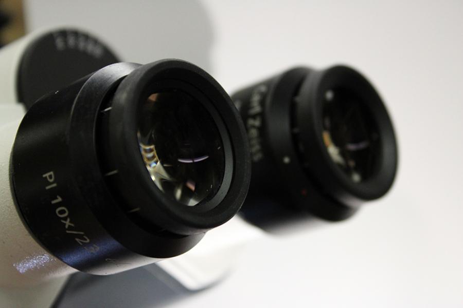
Our imaging facility contains microscopes for brightfield, fluorescence, slide scanning, super-resolution, live cell imaging and analysis. With these versatile imaging tools, our facility is equipped to support a wide array of research needs, from basic biological studies to complex, multi-dimensional analyses.
Training is mandatory for all users. To request training please contact us. We will then arrange a time for training sessions. The length of training is dependent on the user’s previous experience.
It is up to Imaging Facility staff and the department head/platform director to decide whether the user has demonstrated adequate understanding of the instrument to be granted unsupervised access.
Upon completion of training, users must pass a standard practical comprehension test to be granted full privileges including swipe card access.
New users
All new users must contact histology staff to arrange necessary training and initiate fee payments when applicable. This ensures safe and appropriate use of the equipment.
Booking time on instruments
To keep equipment accessible for all, follow these guidelines for booking time:
Facility use
Maintaining a clean and safe environment is essential. Follow these rules when using the facility:
Data storage and file transfer
To protect user data, adhere to these data storage guidelines:
Problems
Immediate reporting of problems ensures a smooth operation for all users. Please notify histology staff of any issues, including supply depletion (oil, lens paper, cleaning solution), software issues, burnt-out bulbs or filaments, or other equipment-related concerns.
After hours / weekends
If problems arise after hours, on weekends, or on holidays, follow these steps: shut down the equipment, place a sign indicating it should not be used, and contact histology staff at histology_services@umanitoba.ca. For emergencies, call 555 for security or 911.
Our imaging and analysis facility provides a variety of equipment tailored to meet diverse research needs, including:
The imaging facility operates on a cost-recovery basis, with all fees contributing to the regular maintenance, repair, and necessary upgrades of the equipment.
UM community rates:
External users rates:
Training rates:
For Department of Human Anatomy and Cell Science research groups requiring more than 30 hours of imaging, an annual fee of $1,500 may apply for unlimited access.
Note: The ELYRA PS.1 system must be operated by facility staff. It is billed at $75/hour for UM community members and $120/hour for external users.
All sample submissions must include the completed forms and the signed estimate.
While forms are available on our website, our team is also available to assist with form completion during the consultation process.
To request services for histology, electron microscopy, or imaging, please ensure you complete the following required forms:
We kindly request that you acknowledge the contributions of the Electron Microscopy Core and Histology Services in any resulting publications. Please use the following statement in your acknowledgments:
"The authors acknowledge the Transmission Electron Microscopy Core Platform and Histology Service Lab at the Department of Human Anatomy and Cell Science, Rady Faculty of Medicine, at the University of Manitoba."
We are committed to helping you showcase your research. If your publications include acknowledgments of the Transmission Electron Microscopy Core Platform and Histology Service Lab, we invite you to feature them on our website. To request this, please contact the Transmission Electron Microscopy Core Platform and Histology Service Lab.
Dr. Sabine Hombach-Klonisch
sabine.hombach-klonisch@umanitoba.ca
Dr. Thomas Klonisch
thomas.klonisch@umanitoba.ca
Dana Henderson
dana.henderson@umanitoba.ca
Farhana Begum
farhana.begum@umanitoba.ca
Nimrat Kaur
Nimrat.kaur@umanitoba.ca
Danyyl Ipolitov
Danyyl.Ipolitov@umanitoba.ca
Histology Services, Imaging Facility, Electron Microscopy Platform
Room 119, 745 Bannatyne Avenue
Basic Medical Science Building
University of Manitoba (Bannatyne campus)
Winnipeg, MB R3E 0J9