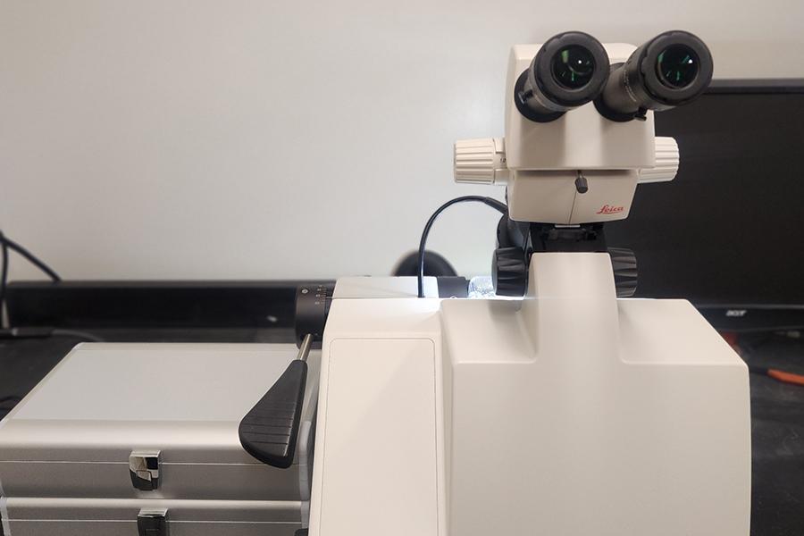
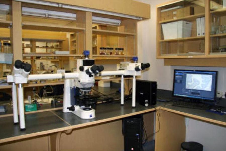
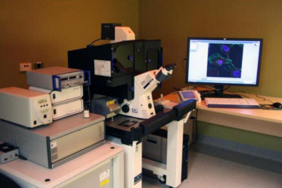
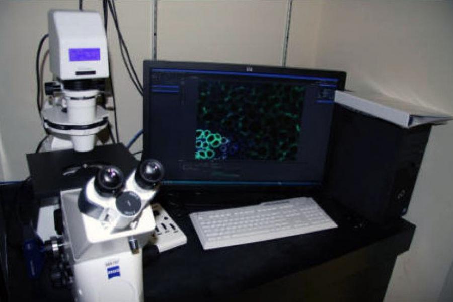
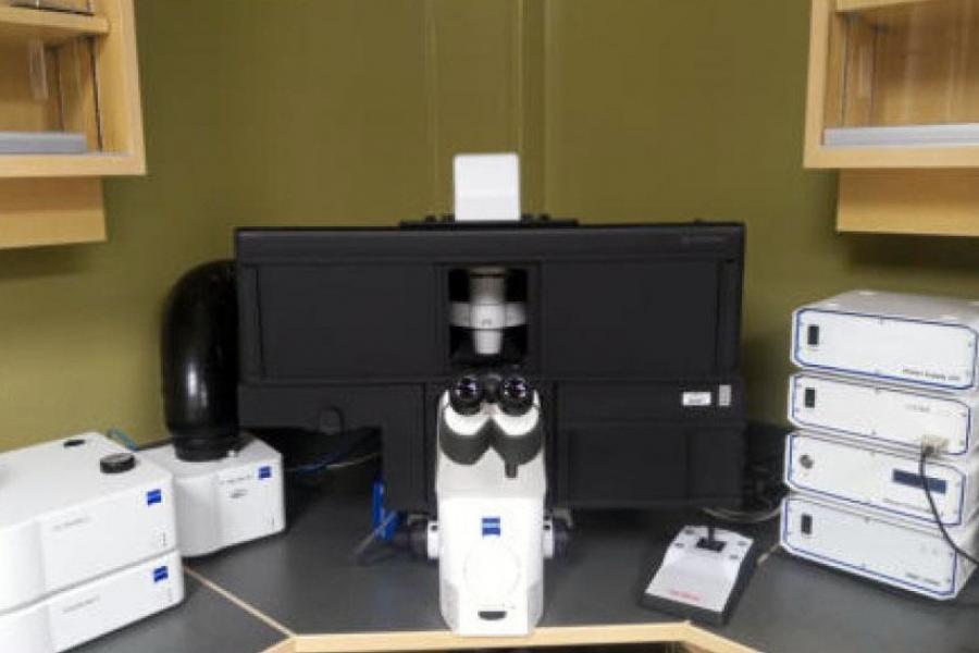
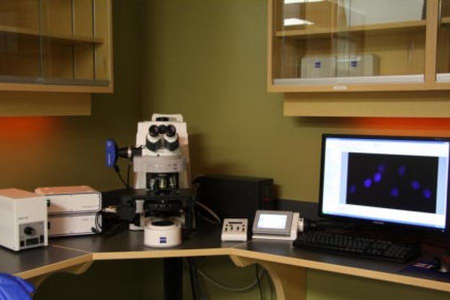
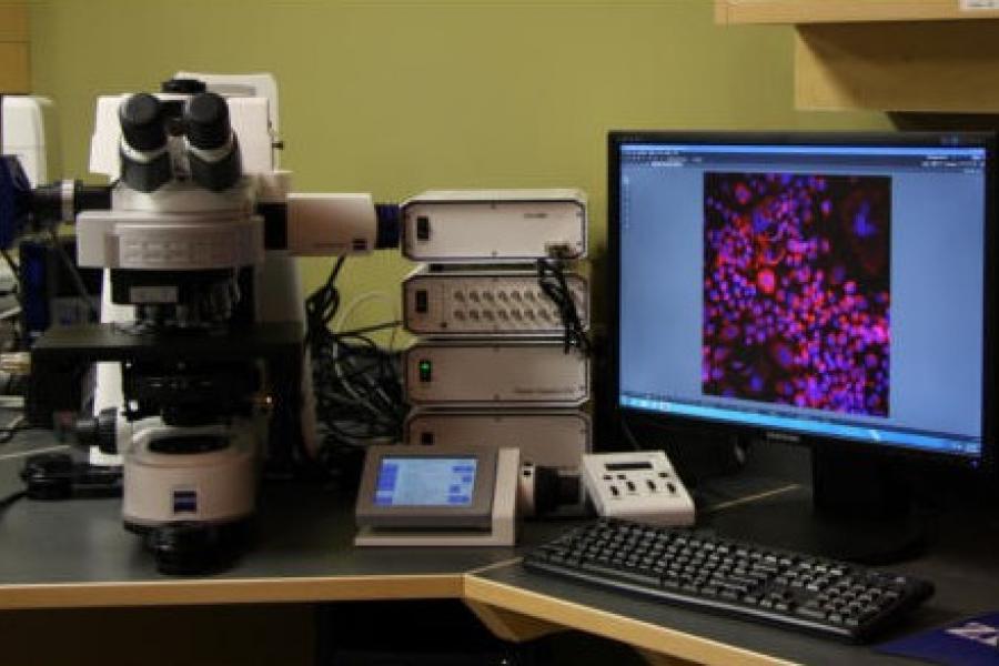
The Zeiss Imager A2 offers advanced imaging capabilities with dual observation arms, precise objective control, and powerful analysis tools.
Below are the key features available for use:
Hardware
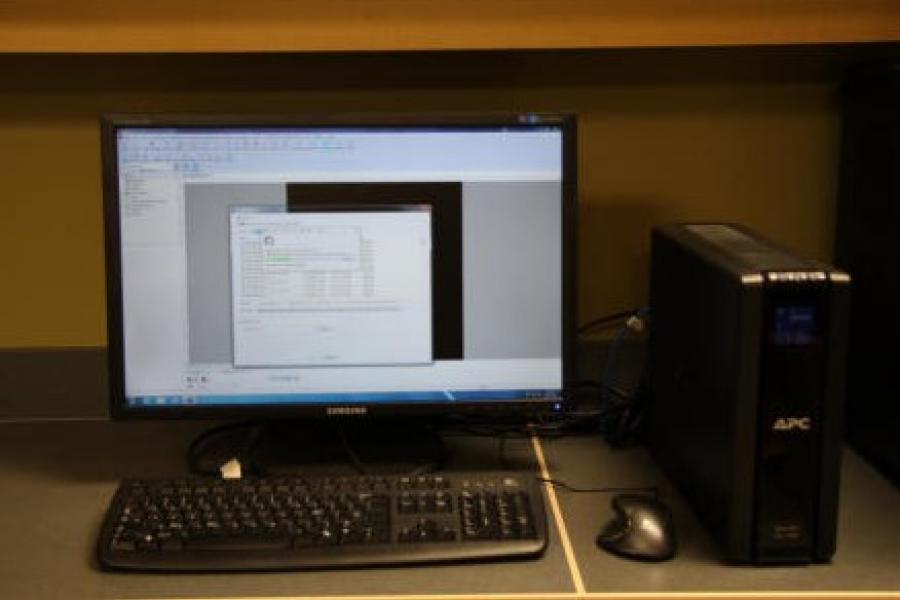
- Computer: HP Z2 (2019), designed to support complex imaging software.
- Dual observation arms: Allow multiple users to observe the sample simultaneously.
Software
The following software is available to support your image analysis and processing:
- ZEN 3 Pro: Offers a range of advanced tools for analyzing and enhancing your images, including:
- Colocalization: Analyzes the overlap of different fluorescent markers to study molecular interactions.
- Extended depth of focus: Combines multiple focal planes into a single image with extended depth for clearer results.
- Time lapse: Records images over time to observe dynamic changes in your sample.
- ZEN Blue Image analysis: Provides digital analysis to extract quantifiable data from your images.
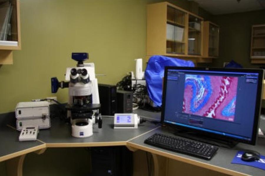
Objective control
The microscope features manual control for the objective and stage for precise positioning:
Stage control
- Manual stage control: X, Y, Z axes.
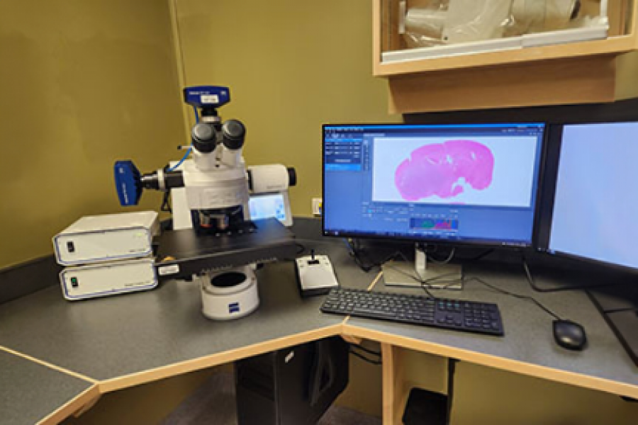
Objectives
The Zeiss Imager A2 is equipped with the following high-quality objectives:
- Zeiss 1.25X/0,03 ∞/0,17 – EC Plan-Neofluar
- Zeiss 5X/0,16 Ph 1 ∞/0,17 – EC Plan-Neofluar
- Zeiss 10X/0,3 ∞/- – EC Plan-Neofluar
- Zeiss 20X/0,3 ∞/0,17 – EC Plan-Neofluar
- Zeiss 40X/0,6 Korr ∞/0-1.5 – LD Plan-Neofluar
- Zeiss 100X/1,3 oil Ph 3 ∞/0,17 – EC Plan-Neofluar
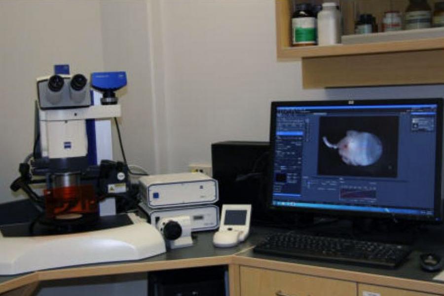
Camera
The system is equipped with two cameras for capturing detailed images:
- AxioCam IcC 5: Colour camera for high-quality imaging.
- AxioCam 208: Colour camera for enhanced image clarity.