Equipment
On this page
Use the links below to jump to the section you're looking for.
-
-
Fluorescence-activated cell sorting
Fluorescence microscopy EVOS Cell Imaging System
-
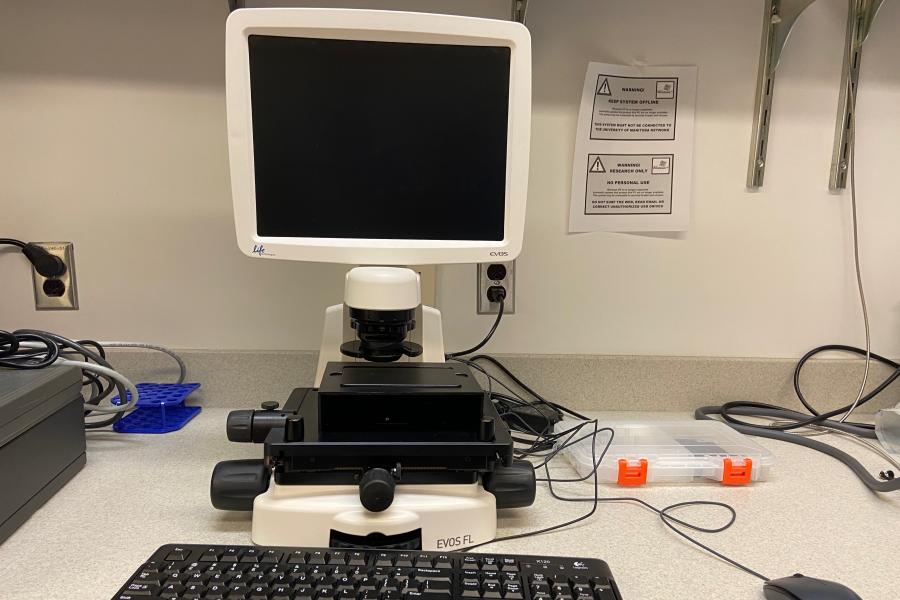
The EVOS Cell Imaging System is a versatile and user-friendly fluorescence microscope designed for rapid imaging of protein expression in live cells. With its ability to image cells while still in tissue culture vessels, it maintains the cells in a viable, sterile state, allowing them to be returned to the incubator after imaging. This system offers multi-colour imaging and supports various sample formats, including chamber slides, regular glass slides, multi-well plates, and tissue culture dishes of different sizes.
Location:
Room 466, Apotex Centre
University of Manitoba (Bannatyne campus) -
Additional details
The EVOS Cell Imaging System offers a flexible, easy-to-use platform for fluorescence microscopy, with features that help streamline your imaging workflow while preserving the viability of your cells. Its wide range of objective lenses, multi-colour imaging capabilities, and compatibility with various sample formats make it a valuable tool for researchers looking for reliable and efficient imaging solutions.
Key features
Light source
- LED light: Provides consistent and bright illumination for a variety of fluorophores, offering long-lasting, stable performance.
Objectives
- 4x, 10x, 20x objectives: Flexible magnification options for a range of imaging needs, from broader views to high-resolution detailed imaging.
Stage accommodation
- Compatible with:
- Regular glass or plastic slides
- Chamber slides
- Multi-well tissue culture plates
- Tissue culture dishes of varying diameters
Channels
- FITC, GFP, Alexa488: For green fluorescence imaging
- Cy3, Rhodamine, Alexa555/568/594: For red fluorescence imaging
- Cy5, APC, Alexa647: For far-red fluorescence imaging
- DAPI: For blue fluorescence (nuclear imaging)
- Bright field: For imaging samples without fluorescence
Fluorescence-activated cell sorting BD FACSMelody
-
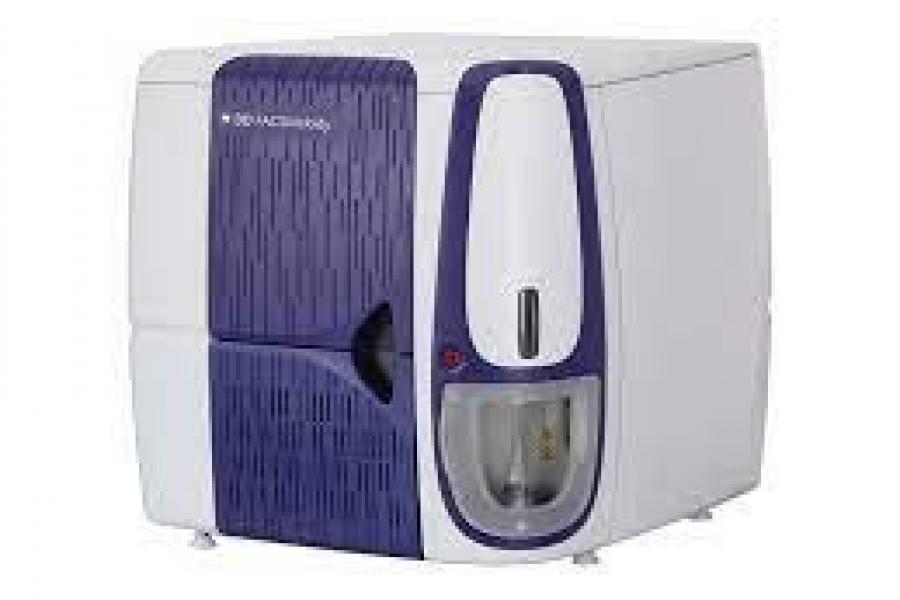
This compact yet powerful instrument, capable of detecting up to 10 parameters and 8 colors simultaneously, makes it possible to identify and isolate even the rarest cell population you are interested in. Samples can be sorted at single cell-basis with sample agitation and temperature control into two or four tubes (5mL round bottom tube to 1.0mL eppendorf), multi-well plates or microscope slides. Our Melody system is housed in a Class-II Type A2 bio-safety cabinet to ensure that samples remain sterile during sorting process. It is capable of accommodating most cell types with its 70, 85 and 100 micron nozzles.
Location
Room 451 (level 2+), Apotex Center
University of Manitoba (Bannatyne Campus) -
This instrument is inside the BSL2+ lab, so only trained users who are on the permit of that room is allowed to stay and work in the room. Those dropping off the samples need to leave the room as soon as they confirm their sample and gating with Christine Zhang.
Additional details
Key features
3 lasers
- 20mW 488nm direct diode blue laser,
- 20mW 405nm direct diode violet laser, and
- 50mW 561nm Solid State violet laser.
17 parameters
- 1-2 Six fluorescent detectors off 488nm,
- 3-4. Three fluorescent detectors off 405nm,
- 5-8. Six fluorescent parameters off 405nm,
- 9. Forward scatter, and
- 10. Side scatter.
| # | Laser | Mirror/Filter | Detection Range | Fluorochrome |
|---|---|---|---|---|
| 1 | blue (488nm) | 507LP, 527/32 | 511-543 | FITC, GFP, Alexa488 |
| 2 | 640LP, 670/30 | 655-685 | PerCP, PE-Cy5, PerCP-Cy5.5, 7-AAD | |
| 3 | violet (405nm) | N/A, 448/45 | 426-470 | V450, BV421, PB, Dapi |
| 4 | 500LP, 528/45 | 506-550 | V500, BV510, AmCyan | |
| 5 | yellow/green (561nm) | 582LP, 582/15 | 575-589 | PE, dsRed |
| 6 | 605LP, 613/18 | 604-622 | mCherry, PE-CF594, PE-TR, PI | |
| 7 | 665LP, 697/58 | 668-726 | PE-Cy5, PE-Cy5.5 | |
| 8 | 752LP, 783/56 | 755-811 | PE-Cy7 |
Acquisition/analysis software
- FACSChorus
Fluorescence-activated cell sorting FACSAria-III
-
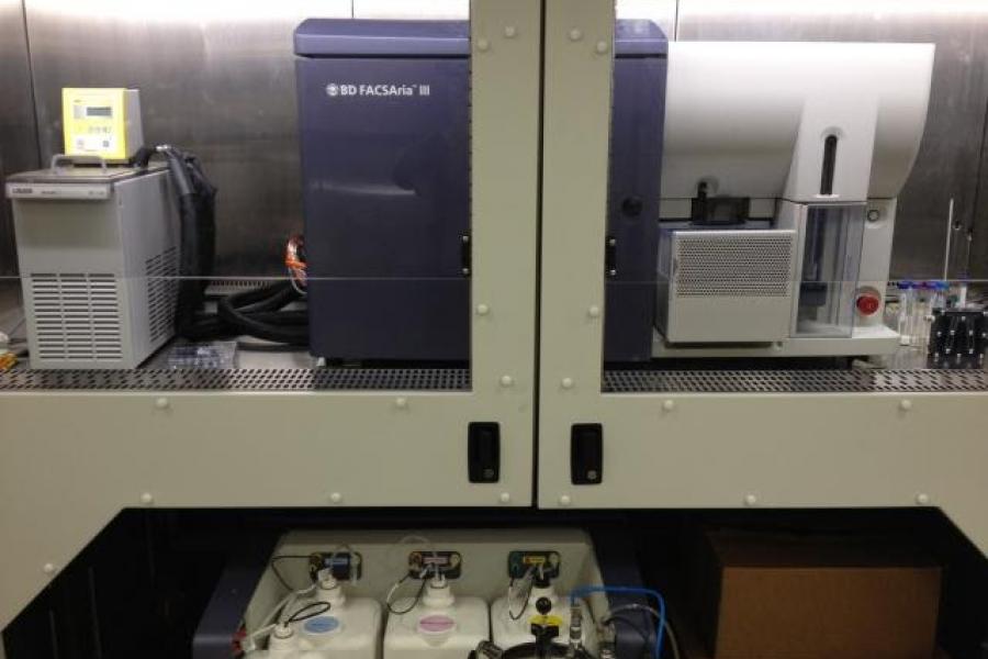
The FACSAria-III is an advanced fluorescence-activated cell sorting (FACS) system that sorts and isolates cell populations with precision, detecting multiple parameters and colours. It allows single-cell sorting with options for agitation and temperature control, making it ideal for diverse research applications. The system accommodates various sample formats, including 15mL centrifuge tubes, 1.0mL microtubes, multi-well plates, and slides. Equipped with an Aerosol Management Option (AMO) for safety and housed in a Class-II Type A2 biosafety cabinet, it ensures sterile conditions during sorting.
Location:
Room 462, Apotex Centre
University of Manitoba (Bannatyne campus) -
Additional details
The FACSAria-III is an essential tool for researchers who need precise, high-throughput cell sorting with multiple parameters and fluorescence detection. Whether isolating rare cell populations or sorting complex samples, this system provides the flexibility, safety, and performance required for cutting-edge research. With advanced features such as aerosol management, biosafety containment, and multiple sorting options, it is the ideal choice for a wide range of cell biology applications.
Key features
Lasers
The FACSAria-III system is equipped with three lasers for fluorescence detection:
- 488nm blue laser: For a wide range of fluorophores, including FITC, GFP, and Alexa488.
- 633nm HeNe red laser: Provides sensitive detection for red fluorescent proteins, including APC, Alexa647, and Cy5.
- 405nm violet laser: Enables detection of UV-excitable fluorophores like Pacific Blue, DAPI, and BrilliantViolet dyes.
Nozzle sizes
- 70, 85, and 100 microns: These nozzles accommodate most cell types, providing flexibility for sorting a wide range of sample sizes.
Sample sorting
- Sort samples at a single-cell level with precise control over temperature and agitation.
- Sort into various containers, including:
- 15mL centrifuge tubes
- 1.0mL microtubes
- Multi-well plates
- Microscope slides
Aerosol Management Option (AMO)
- A safety feature that traps aerosolized particles, reducing the risk of contamination and ensuring safe operation in high-risk experiments.
Bio-safety containment
- The FACSAria-III system is housed in a Class-II Type A2 biosafety cabinet, ensuring that your samples remain sterile during the sorting process.
Fluorescent detection channels
The FACSAria-III can detect up to 17 parameters using 15 different fluorochromes. These are distributed across the three lasers and multiple detection channels, allowing for complex multi-parameter analysis.
Here’s a breakdown of the detection ranges and fluorochromes:
| # | Laser | Mirror/Filter | Detection Range | Fluorochrome |
|---|---|---|---|---|
| 1 | blue (488nm) | 502LP, 530/30 | 515-545 | FITC, GFP, Alexa488 |
| 2 | 556LP, 585/42 | 564-606 | PE, dsRed | |
| 3 | 610LP, 616/23 | 610-627 | PE-Texas Red, PI (live) | |
| 4 | 640LP, 670/30 | 655-685 | PerCP, PE-Cy5, 7-AAD | |
| 5 | 685LP, 695/40 | 685-715 | PE-Cy5.5, PerCP-Cy5.5 | |
| 6 | 735LP, 780/60 | 750-810 | PE-Cy7, PE-Alexa750 | |
| 7 | red (633nm) | N/A, 660/20 | 650-670 | APC, Alexa647, Cy5 |
| 8 | 690LP, 730/45 | 708-752 | Alexa700 | |
| 9 | 735LP, 780/60 | 750-810 | APC-cy7, APC-H7 | |
| 10 | violet (405nm) | N/A, 450/40 | 430-470 | Pacific blue, Dapi, V450, BV421 |
| 11 | 502LP, 510/50 | 502-545 | AmCyan, CFP, V500, BV510 | |
| 12 | 545LP, 560/20 | 550-570 | BrilliantViolet570 | |
| 13 | 595LP, 610/20 | 578-592 | BrilliantViolet605 | |
| 14 | 630LP, 660/20 | 650-670 | BrilliantViolet650 | |
| 15 | 690LP, 710/50 | 685-735 | BrilliantViolet711 |
Flow cytometry cell analysis 4L YG Aurora
-
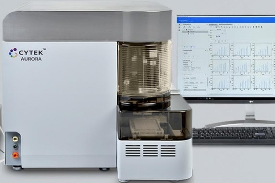
The Cytek Aurora spectral flow cytometry analyzer is equipped with 4 lasers and capable of detecting up to 48 fluorescent parameters, giving researchers more flexibility and room to easy design a large panel of markers of over 20 colors. Besides the standard forward and side scatter channels off from blue laser, it also comes with side scatter channel off from 100mw 405nm violet laser, allowing you to detect small particles as small as nearing 100nm.
Other than the conventional individual 75mm round bottom tube loading, Aurora also comes with high throughput Automated Sample Loader, compatible for 96 well plates, 96 deep-well plates and 40 tube racks to streamline the acquisition work.
Location:
Room 466, Apotex Centre
University of Manitoba (Bannatyne Campus) -
Additional details
Key features
Sample handling
- Semi-automatic Single Tube Loading with 12 x 75 mm or 1.2 mL tubes
- High Throughput Automated Sample Loader unit for 96 well plates, 96 deep-well plates and 40 tube racks
4 lasers
- 50mW 488nm Solid State blue laser
- 80mW 640nm Solid State red laser
- 100mW 405nm Solid State violet laser
- 50mW 561nm Solid State yellow-green laser
51 parameters
- 1-14: fourteen fluorescent detectors off blue laser
- 15-22: eight fluorescent detectors off red laser
- 23-38: sixteen fluorescent detectors off violet laser
- 38-48: ten fluorescent detectors off yellow-green laser
- 49: Blue Laser Forward scatter
- 50: Blue Laser Side scatter
- 51: Violet Laser Side scatter
| Channel | Laser | Mirror/Filter | Detection Range | Fluorochrome |
|---|---|---|---|---|
| B1 | blue (488nm) | 508/20 | 498-518 | BB515, VioBright B515, Vio B515, NovaFluor Blue 510, eGFP, ZsGreen |
| B2 | 525/17 | 516-534 | FITC, AF488, VioBright FITC, KIRAVIA Blue 520, CFSE | |
| B3 | 542/17 | 533-551 | RB545, AF532, Spark Blue 550, NovaFluor Blue 530, NovaFluor Blue 555, eYFP, mVenus | |
| B4 | 581/19 | 571-591 | StarBright Blue 580, Spark Blue 574 | |
| B5 | 598/20 | 588-608 | NovaFluor Blue 585 | |
| B6 | 615/20 | 605-625 | BB630, StarBright Blue 615, NovaFluor Blue 610 30S, NovaFluor Blue 610-70S | |
| B7 | 661/17 | 652-670 | BB670 | |
| B8 | 679/18 | 670-688 | PerCP, StarBright Blue 675, NovaFluor Blue 660 120S | |
| B9 | 697/19 | 687-707 | PerCP-Cy5.5, BB700, StarBright Blue 700 | |
| B10 | 717/20 | 707-727 | PerCP-eF710, PerCP-Vio700 | |
| B11 | 738/21 | 727-748 | ||
| B12 | 760/23 | 748-772 | BB755, StarBright Blue 765 | |
| B13 | 783/23 | 772-795 | ||
| B14 | 812/24 | 795-829 | RB780, BB790, StarBright Blue 810 | |
| R1 | red (640nm) | 661/17 | 652-670 | APC, STYOX Red, Cy5 |
| R2 | 679/18 | 670-688 | AF647, eF660, Vio R667, Vio Bright R667, NovaFluor Red 660, NovaFluor Blue 660 40S, miRFP670 | |
| R3 | 697/19 | 688-707 | NovaFluor Red 700, Alexa Fluor 660, Spark NIR 685, NovaFluor Red 685, miRFP | |
| R4 | 717/20 | 707-727 | AF700, Red 718, APC-R700, Vio Bright R720, redFluor710, NovaFluor 710 | |
| R5 | 738/21 | 727-749 | NovaFluor Red 725, Spark Red 718 | |
| R6 | 760/23 | 749-772 | ||
| R7 | 783/23 | 772-795 | APC-eF780, APC-Cy7, APC-H7, APC-Vio770, APC-Fire 750, NovaFluor Red 755, GhostDye Red 780 | |
| R8 | 812/34 | 795-829 | APC-Fire810 | |
| V1 | Violet (405nm) | 428/15 | 420-436 | BV421 |
| V2 | 443/15 | 436-451 | SuperBright 436, Alexa Fluor 405, Vio Bright V423, Spark Violet 423, StarBright Violet 440 | |
| V3 | 458/15 | 451-466 | BeF450, V450, Pacific Blue, vF450, VioBlue, eBFP, eBFP2 | |
| V4 | 473/15 | 466-481 | SYTOX Blue | |
| V5 | 508/20 | 498-518 | BV480, V500, Spark Violet 500, StarBright Violet 475 | |
| V6 | 525/17 | 518-534 | StarBright Violet 515, mAmetrine | |
| V7 | 542/17 | 534-551 | BV510. Spark Violet 538, VioGreen, Live/Dead Fixable Aqua | |
| V8 | 581/19 | 571-591 | BV570, StarBright Violet 570, Pacific Orange | |
| V9 | 598/20 | 588-608 | ||
| V10 | 615/20 | 605-625 | BV605, SuperBright 600, StarBright Violet 610, mKeima | |
| V11 | 664/27 | 650-678 | BV650 ,SuperBright 645, StarBright Violet 670 | |
| V12 | 692/28 | 678-706 | ||
| V13 | 720/29 | 706-735 | BV711, SuperBright 702, StarBright Violet 710 | |
| V14 | 750/30 | 735-765 | BV750, StarBright Violet 760 | |
| V15 | 780/30 | 765-795 | BV786, BV785, SuperBright 780, StarBright Violet 790 | |
| V16 | 812/34 | 795-829 | ||
| YG1 | Yellow-Green laser (561nm) | 577/20 | 567-587 | PE, Alexa Fluor 555, StarBright Yellow 575, NovaFluor Yellow 570, Spark YG 570, Spark YG 581, tdTomato, RFP, DsRed |
| YG2 | 598/20 | 588-608 | RY586, NovaFluor Yellow 590, Spark YG 593 | |
| YG3 | 615/20 | 605-625 | PE-CF594, PE-Texas Red, PE-eF610, PE-Dazzle 594, PE-Vio615, StarBright Yellow 605, Alexa Fluor 594, NovaFluor Yellow 610, mCherry | |
| YG4 | 661/17 | 652-670 | PI, PE-Fire 640, StarBright Yellow 665, NovaFluor Yellow 660, 7-AAD | |
| YG5 | 679/18 | 670-688 | PE-Cy5 | |
| YG6 | 697/19 | 670-688 | NovaFluor Yellow 690 | |
| YG7 | 720/29 | 705-735 | PE-Cy5.5, PE-Fire 700, StarBright Yellow 720, NovaFluor Yellow 700, NovaFluor Yellow 730 | |
| YG8 | 750/30 | 735-765 | ||
| YG9 | 780/30 | 765-795 | PE-Cy7, PE-Vio770, StarBright Yellow 800, NovaFluor Yellow 755 | |
| YG10 | 812/34 | 795-829 | PE-Cy7, PE-Fire810 |
Acquisition/analysis software
- SpectroFlo
Notes
- How to read longpass filter detection range:
- e.g., 502LP—wavelenge Longer than 502nm will be allowed to Pass through
- How to read bandpass filter detection range:
- e.g., 530/30 = 530nm ± (30/2)nm = 530nm ± 15nm = from 515nm to 545nm
Flow cytometry cell analysis 5L Aurora
-
The Cytek Aurora spectral flow cytometry analyzer is equipped with 5 lasers and capable of detecting up to 64 fluorescent parameters, giving researchers more flexibility and room to easy design a large panel of markers of over 20 colors. Besides the standard forward and side scatter channels off from blue laser, it also comes with side scatter channel off from 100mw 405nm violet laser.
Other than the conventional individual 75mm round bottom tube loading, Aurora also comes with high throughput Automated Sample Loader, compatible for 96 well plates, 96 deep-well plates and 40 tube racks to streamline the acquisition work.
Location:
Room 531, Basic Medical Science Building
University of Manitoba (Bannatyne Campus) -
Additional details
Key features
Sample handling
- Semi-automatic Single Tube Loading with 12 x 75 mm or 1.2 mL tubes
- High Throughput Automated Sample Loader unit for 96 well plates, 96 deep-well plates and 40 tube racks
5 lasers
- 50mW 488nm Solid State blue laser,
- 80mW 640nm Solid State red laser
- 100mW 405nm Solid State violet laser.
- 50mW 561nm Solid State yellow-green laser
- 20mW 355nm Solid State UV laser
67 parameters
- 1-14: fourteen fluorescent detectors off blue laser
- 15-22: eight fluorescent detectors off red laser
- 23-38: sixteen fluorescent detectors off violet laser
- 38-48: ten fluorescent detectors off yellow-green laser
- 49-64: sixteen fluorescent detectors off UV laser
- 65: Blue Laser Forward scatter
- 66: Blue Laser Side scatter
- 67: Violet Laser Side scatter
| Laser | Mirror/Filter | Detection Range | Fluorochrome |
|---|---|---|---|
| B1 (blue 488nm) | 508/20 | 498-518 | BB515, VioBright B515, Vio B515, NovaFluor Blue 510, eGFP, ZsGreen |
| B2 | 525/17 | 516-534 | FITC, AF488, VioBright FITC, KIRAVIA Blue 520, CFSE |
| B3 | 542/17 | 533-551 | RB545, AF532, Spark Blue 550, NovaFluor Blue 530, NovaFluor Blue 555, eYFP, mVenus |
| B4 | 581/19 | 571-591 | StarBright Blue 580, Spark Blue 574 |
| B5 | 598/20 | 588-608 | NovaFluor Blue 585 |
| B6 | 615/20 | 605-625 | BB630, StarBright Blue 615, NovaFluor Blue 610 30S, NovaFluor Blue 610-70S |
| B7 | 661/17 | 652-670 | BB670 |
| B8 | 679/18 | 670-688 | PerCP, StarBright Blue 675, NovaFluor Blue 660 120S |
| B9 | 697/19 | 687-707 | PerCP-Cy5.5, BB700, StarBright Blue 700 |
| B10 | 717/20 | 707-727 | PerCP-eF710, PerCP-Vio700 |
| B11 | 738/21 | 727-748 | |
| B12 | 760/23 | 748-772 | BB755, StarBright Blue 765 |
| B13 | 783/23 | 772-795 | |
| B14 | 812/24 | 795-829 | RB780, BB790, StarBright Blue 810 |
| R1 (red 640nm) | 661/17 | 652-670 | APC, STYOX Red, Cy5 |
| R2 | 679/18 | 670-688 | AF647, eF660, Vio R667, Vio Bright R667, NovaFluor Red 660, NovaFluor Blue 660 40S, miRFP670 |
| R3 | 697/19 | 688-707 | NovaFluor Red 700, Alexa Fluor 660, Spark NIR 685, NovaFluor Red 685, miRFP |
| R4 | 717/20 | 707-727 | AF700, Red 718, APC-R700, Vio Bright R720, redFluor710, NovaFluor 710 |
| R5 | 738/21 | 727-749 | NovaFluor Red 725, Spark Red 718 |
| R6 | 760/23 | 749-772 | |
| R7 | 783/23 | 772-795 | APC-eF780, APC-Cy7, APC-H7, APC-Vio770, APC-Fire 750, NovaFluor Red 755, GhostDye Red 780 |
| R8 | 812/34 | 795-829 | APC-Fire810 |
| V1 (violet 405nm) | 428/15 | 420-436 | BV421 |
| V2 | 443/15 | 436-451 | SuperBright 436, Alexa Fluor 405, Vio Bright V423, Spark Violet 423, StarBright Violet 440 |
| V3 | 458/15 | 451-466 | BeF450, V450, Pacific Blue, vF450, VioBlue, eBFP, eBFP2 |
| V4 | 473/15 | 466-481 | SYTOX Blue |
| V5 | 508/20 | 498-518 | BV480, V500, Spark Violet 500, StarBright Violet 475 |
| V6 | 525/17 | 518-534 | StarBright Violet 515, mAmetrine |
| V7 | 542/17 | 534-551 | BV510, Spark Violet 538, VioGreen, Live/Dead Fixable Aqua |
| V8 | 581/19 | 571-591 | BV570, StarBright Violet 570, Pacific Orange |
| V9 | 598/20 | 588-608 | |
| V10 | 615/20 | 605-625 | BV605, SuperBright 600, StarBright Violet 610, mKeima |
| V11 | 664/27 | 650-678 | BV650, SuperBright 645, StarBright Violet 670 |
| V12 | 692/28 | 678-706 | |
| V13 | 720/29 | 706-735 | BV711, SuperBright 702, StarBright Violet 710 |
| V14 | 750/30 | 735-765 | BV750, StarBright Violet 760 |
| V15 | 780/30 | 765-795 | BV786, BV785, SuperBright 780, StarBright Violet 790 |
| V16 | 812/34 | 795-829 | |
| YG1 (yellow-green 561nm) | 577/20 | 567-587 | PE, Alexa Fluor 555, StarBright Yellow 575, NovaFluor Yellow 570, Spark YG 570, Spark YG 581, tdTomato, RFP, DsRed |
| YG2 | 598/20 | 588-608 | RY586, NovaFluor Yellow 590, Spark YG 593 |
| YG3 | 615/20 | 605-625 | PE-CF594, PE-Texas Red, PE-eF610, PE-Dazzle 594, PE-Vio615, StarBright Yellow 605, Alexa Fluor 594, NovaFluor Yellow 610, mCherry |
| YG4 | 661/17 | 652-670 | PI, PE-Fire 640, StarBright Yellow 665, NovaFluor Yellow 660, 7-AAD |
| YG5 | 679/18 | 670-688 | PE-Cy5 |
| YG6 | 697/19 | 670-688 | NovaFluor Yellow 690 |
| YG7 | 720/29 | 705-735 | PE-Cy5.5, PE-Fire 700, StarBright Yellow 720, NovaFluor Yellow 700, NovaFluor Yellow 730 |
| YG8 | 750/30 | 735-765 | |
| YG9 | 780/30 | 765-795 | PE-Cy7, PE-Vio770, StarBright Yellow 800, NovaFluor Yellow 755 |
| YG10 | 812/34 | 795-829 | PE-Cy7, PE-Fire810 |
| UV1 (UV 355nm) | 373/15 | 365-380 | Spark UV 387 |
| UV2 | 388/15 | 381-395 | BUV395, StarBright UV 400 |
| UV3 | 428/15 | 421-435 | |
| UV4 | 443/15 | 436-450 | FVS440UV |
| UV5 | 458/15 | 451-465 | |
| UV6 | 473/15 | 466-480 | Alexa Fluor 350, StarBright UV 445, Live/Dead Blue |
| UV7 | 514/28 | 500-528 | BUV496, StarBright UV 510, DAPI |
| UV8 | 542/28 | 528-556 | |
| UV9 | 582/31 | 566-598 | BUV563, StarBright UV 575 |
| UV10 | 613/31 | 598-629 | BUV615, StarBright UV 605 |
| UV11 | 664/27 | 650-678 | BUV661, StarBright UV 665 |
| UV12 | 692/28 | 678-706 | |
| UV13 | 720/29 | 706-734 | |
| UV14 | 750/30 | 735-765 | BUV737, StarBright UV 740 |
| UV15 | 780/30 | 765-795 | |
| UV16 | 812/34 | 795-829 | BUV805, StarBright UV 795 |
Acquisition/analysis software
- SpectroFlo
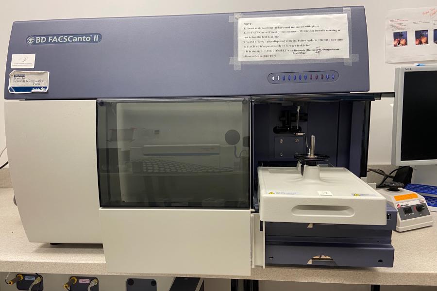
Flow cytometry cell analysis FACSCanto II
-
The FACSCanto II is a powerful flow cytometry system designed for both clinical and research applications. Capable of detecting up to 10 parameters and eight colours simultaneously, it allows researchers to perform detailed cell analysis with high precision. The newly designed loader carousel enables automatic loading of up to 40 12x75mm tubes, reducing hands-on time and improving result reliability.
Location:
Room 466, Apotex Centre
University of Manitoba (Bannatyne Campus) -
Additional details
The FACSCanto II is ideal for researchers looking for efficiency and precision. Its semi-automatic loading system minimizes operator intervention, saving time and reducing potential errors. With its three lasers and a wide range of fluorescence detection options, it supports a variety of experiments requiring multiple parameters. The system is designed to handle a large volume of samples, making it a valuable tool for both routine and complex analyses.
Key features
Sample handling
- Semi-automatic single tube loading with 12 x 75 mm tubes
- Can automatically load up to 40 tubes without operator intervention
Lasers
- 488nm Solid State blue laser (OmW)
- 633nm HeNe red laser (7mW)
- 405nm Solid State violet laser (OmW)
Detection parameters
- 10 parameters total:
- 1-4: Four fluorescent detectors off the 488nm laser
- 5-6: Two fluorescent detectors off the 633nm laser
- 7-8: Two fluorescent parameters off the 405nm laser
- 9: Forward scatter
- 10: Side scatter
Fluorochrome and filter details
The FACSCanto II offers various filter and mirror options for a wide range of fluorochromes. Here are some of the key channels:
| # | Laser | Mirror/Filter | Detection Range | Fluorochrome |
|---|---|---|---|---|
| 1 | blue (488nm) | 502LP, 530/30 | 515-545 | FITC, GFP, Alexa488 |
| 2 | 556LP, 585/42 | 564-606 | PE, dsRed | |
| 3 | 655LP, 670LP | ≥670 | PerCP, PE-Cy5.5, PI, 7-AAD | |
| 4 | 735LP, 780/60 | 750-810 | PE-Cy7 | |
| 5 | red (633nm) | 660/20 | 650-670 | APC, Alexa647, Cy5 |
| 6 | 735LP, 780/60 | 750-810 | APC-cy7, APC-H7 | |
| 7 | violet (405nm) | 450/50 | 425-475 | Pacific blue, Dapi, V450, BV421 |
| 8 | 502LP, 510/50 | 502-535 | AmCyan, V500, BV510 |
Software
The FACSCanto II uses FACSDiva acquisition and analysis software, which enables easy setup, data collection, and analysis for efficient workflows.
Notes
- Longpass filter detection range: e.g., 502LP—wavelength longer than 502nm will be allowed to pass through.
- Bandpass filter detection range: e.g., 530/30 = 530nm ± (30/2)nm = 530nm ± 15nm = from 515nm to 545nm.
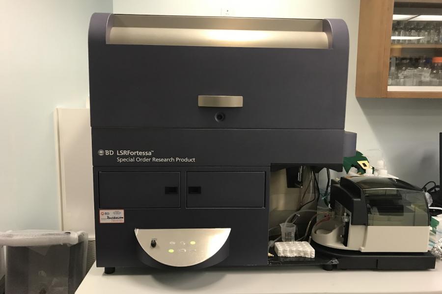
Flow cytometry cell analysis Fortessa
-
The Fortessa is an advanced flow cytometry system designed for high-throughput, multicolour assays. Capable of detecting up to 18 fluorescent parameters, this instrument is ideal for complex cell analysis involving large marker panels. It offers flexibility with both single tube loading and a high throughput sampler unit for multi-well plates, making it suitable for a wide range of research and clinical applications.
Location:
Room 536, Basic Medical Science Building
University of Manitoba (Bannatyne campus) -
Additional details
The Fortessa is perfect for researchers needing detailed multicolour analysis, capable of detecting up to 18 fluorescent parameters. Its high-throughput sampler unit speeds up sample acquisition in multi-well plates, while flexible sample handling options save time. Equipped with five lasers (blue, red, violet, yellow-green, and UV), it supports a wide range of fluorochromes and applications, offering versatility and efficiency for complex experiments.
Key features
Sample handling
- Semi-automatic single tube loading with 12 x 75 mm or 1.2 mL tubes
- High throughput sampler unit for multi-well plates, available in standard and high-throughput modes
Lasers
The Fortessa is equipped with five solid-state lasers to enable broad spectrum fluorescence detection:
- 50mW 488nm solid state blue laser
- 40mW 633nm solid state red laser
- 50mW 405nm solid state violet laser
- 50mW 561nm solid state yellow-green laser
- 20mW 355nm solid state UV laser
Detection parameters
- Blue laser (488nm): Detects up to 2 fluorescent parameters
- Red laser (640nm): Detects up to 3 fluorescent parameters
- Violet laser (405nm): Detects up to 6 fluorescent parameters
- Yellow-green laser (561nm): Detects up to 4 fluorescent parameters
- UV laser (355nm): Detects up to 3 fluorescent parameters
- Forward scatter (FSC) and side scatter (SSC) for cell size and granularity analysis
Fluorochrome and filter details
Each laser has specific filters to detect a wide range of fluorochromes:
| # | Laser | Mirror/Filter | Detection Range | Fluorochrome |
|---|---|---|---|---|
| 1 | blue (488nm) | 505LP, 530/30 | 515-545 | FITC, GFP, Alexa488 |
| 2 | 685LP, 710/50 | 685-735 | PerCP-Cy5.5 | |
| 3 | red (640nm) | 670/14 | 663-667 | APC, Alexa647, Cy5 |
| 4 | 690LP, 730/45 | 708-752 | Alexa700 | |
| 5 | 750LP, 780/60 | 750-810 | APC-cy7, APC-H7 | |
| 6 | violet (405nm) | 450/50 | 425-475 | Pacific blue, Dapi, V450, BV421 |
| 7 | 505LP, 525/50 | 505-550 | AmCyan, CFP, V500, BV510 | |
| 8 | 600LP, 610/20 | 600-620 | BrilliantViolet605 | |
| 9 | 630LP, 670/30 | 655-685 | BrilliantViolet650 | |
| 10 | 690LP, 710/50 | 685-735 | BrilliantViolet711 | |
| 11 | 750LP, 780/60 | 750-810 | BrilliantViolet786 | |
| 12 | yellow-green laser (561nm) | 545LP, 586/16 | 578-594 | PE |
| 13 | 600LP, 610/20 | 600-620 | PE-Taxas Red | |
| 14 | 635LP, 670/30 | 655-685 | PE-Cy5 | |
| 15 | 750LP, 780/60 | 750-810 | PE-Cy7 | |
| 16 | UV laser (355nm) | 379/28 | 365-393 | BUV395 |
| 17 | 450LP, 515/30 | 500-530 | BUV496 | |
| 18 | 690LP, 740/35 | 723-757 | BUV737 |
Software
The Fortessa uses FACSDiva software for acquisition and analysis, offering an intuitive platform for data collection, visualization, and analysis.
Notes
- Longpass filter detection range: A filter like 502LP allows wavelengths longer than 502nm to pass through.
- Bandpass filter detection range: A filter like 530/30 detects wavelengths within 530nm ± 15nm (515-545nm).
Equipment reservation
The Flow Core has moved its instrument booking system to a new platform on SuperSaaS. All current users must create a new account on this platform.
Only users who were previously trained and had access to the old booking calendar are eligible to register.
New users must complete full instrument training before they can sign up or gain access to the new booking system.
To reserve an instrument for flow cytometry analysis or cell sorting, please use our online booking platform.
Reservation policy
All new users must complete the mandatory instrument training before being granted access to our online booking system. Once the training is completed and budget number provided, the core facility supervisor will add you onto the member list of our online booking system. You will then be able to view the calendar and book the flow cytometry analyzers of your choice at the available time.
Please read to following important guidelines when reserving instrument for your use:
- Your online booking calendar ID is meant to be used by you ONLY. Please do not share it with others, especially with the untrained users. You should ALWAYS make reservation on the instruments before using the flow cytometers as they are shared by many research groups.
- There are six instruments housed in our facility (FACSCanto-II, LSR-II, FACSAria-III, Cytoflex LX, LSRFortessa, Zeiss Confocal), each instrument has its own calendar. Please select and reserve the correct instrument you have been trained on from the drop-down list on the upper-right corner.
- Please note that you cannot make changes on the event one hour prior to the scheduled time. The instruments, especially the FACSCanto-II, are used heavily by many users, so be considerate to others and reserve only the length of the time you need. ALWAYS cancel your booking in advance if you cannot make it to your appointment. If cancellation is within the same day of your scheduled time, contact me IMMEDIATELY. This is for emergency only and we do not accept booking cancellation after the scheduled booking has passed.
- Although the calendar only shows time from 7am to 6:30pm by default. Instrument reservation is still required for any usage before 7:00 am or after 6:30 pm. You can expand the calendar to see or make bookings before 7:00 am or after 6:30 pm by clicking the small yellow triangle on the top right corner in the Day Grid View.
- You are granted access to reservation of ONLY the instrument you have been trained on (either FACSCanto-II or LSR-II). Additional training is required if you'd like to use the other instrument. Please contact me first to arrange for a training course before reserving the instrument for use.
- Reservation and operation of the FACSAria-III for cell sorting will be done only by the Core Facility Supervisor. Regular users will be able to view but not to book sorting on the calendar. Please check the calendar for available time slot and contact the facility manager by email to book for a cell sorting service.
Contact us
Flow Cytometry
413 Apotex Center, 750 McDermot Avenue
University of Manitoba
Winnipeg, MB, R3E 0T5, Canada
204-294-0691
204-789-3921