Overview

Examples
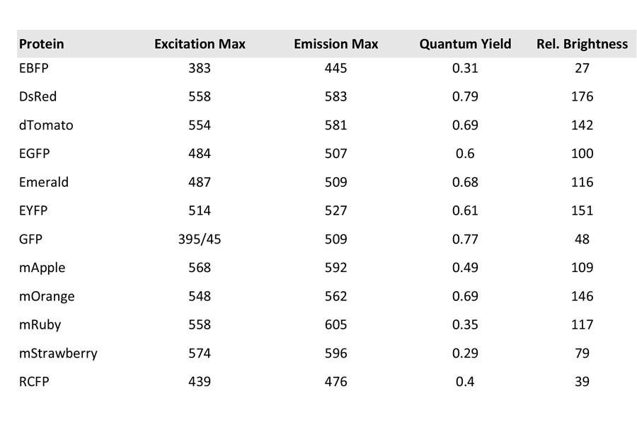
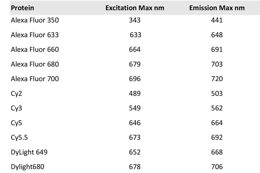
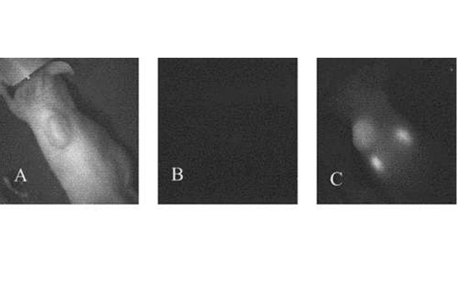
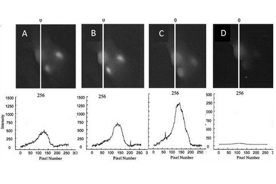
One of the most common uses of optical imaging is preclinical studies of anti-tumour agents. In the example shown here, fluorescence imaging was used to study the binding of a novel antitumour antibody, NovoMab-G2-scFv, to tumour cells. NovoMab-G2-scFv was labelled with Cy5.5.18, a fluorescent dye with an absorption maximum at 670 nm and emission maximum at 720 nm.
Instrumentation
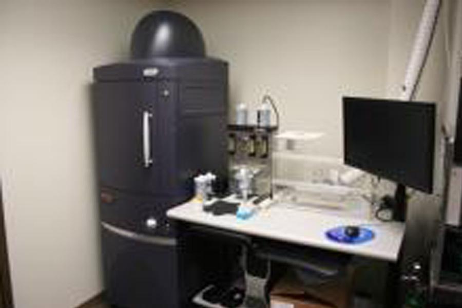
Perkin Elmer/Caliper IVIS Spectrum
Optical imaging is performed using a Perkin Elmer/Caliper IVIS Spectrum optical imaging system.
Additional information
Fluorescence and bioluminescence images are acquired using a high sensitivity, back thinned, back illuminated CCD camera cooled to -90 degrees Celsius.
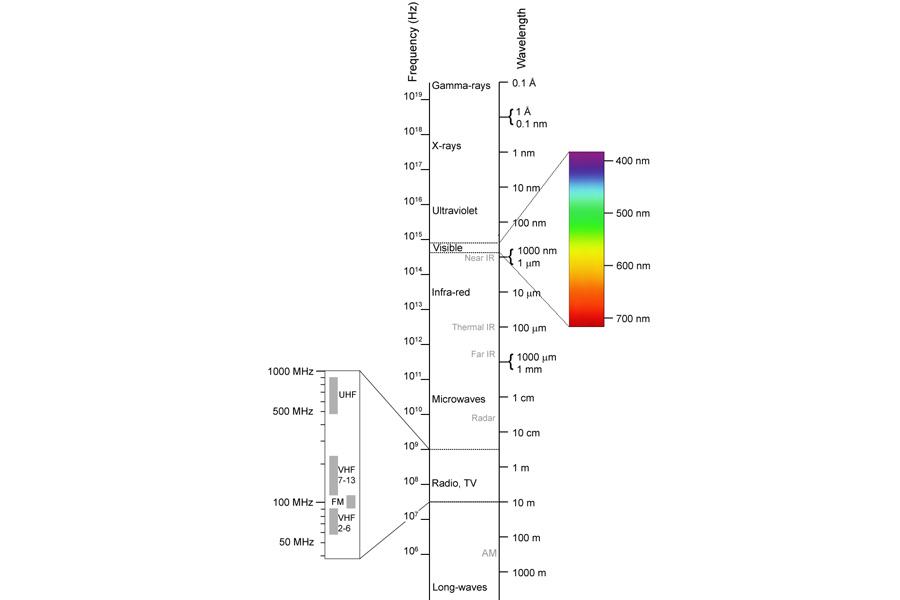
Spectral region selected is enabled by 10 narrow band excitation (430, 465, 500, 535, 570, 606, 640, 675, 710 and 745 nm, 30 nm bandpass) and 18 narrow band emission (500, 520, 540, 560, 580, 600, 620, 640, 660, 680, 700, 720, 740, 760, 780, 800, 820 and 840 nm, 20 nm bandpass) filters, optimized to allow data acquisition from the blue to near infrared wavelength regions (430 nm to 850 nm).
For fluorescence imaging, the IVIS Spectrum allows investigators to use either trans-illumination (from the bottom) or epi-illumination (from the top) to illuminate experimental animals. Using a combination of a structured light source and trans-illumination investigators can perform 3D diffuse fluorescence tomography to determine source localization and concentration.
Four fields of view, (22.5 cm, 12.5 cm, 6.5 cm, and 3.9 cm) are selectable, allowing studies of up to five mice simultaneously or a single sample with high spatial resolution (up to 20 um).
Applications
Applications include:
- Non-invasive imaging of animal models of human disease
- Non-invasive imaging of genetically engineered animals
- Assess efficacy, pharmacokinetics and biodistribution of novel pharmacological agents
- Assess novel drug delivery and gene therapy approaches
- Develop new optical reporters for diagnostic imaging
Resources
Application notes
The materials listed here are available for investigators by request.
Adaptive fluorescence background subtraction
Autoexposure
Background ROI
Determine saturation
DLIT1 setup
DLIT2 topography
DLIT3 reconstruction
Drawing ROIs
FLIT 1 setup
FLIT 2 topography
FLIT 3 reconstruction
FMT imaging techniques
FMT multispecies imaging module (MSIM) for FMT
High resolution images
Image math
Image overlay 2D
Image overlay 3D
Imaging wizard
Load as group
Sending large files for analysis
Spectral unmixing
Subject ROI
Transillumination 1 setup
Transillumination 2 raster scan
Transillumination 3 normalized
Well plate quantification
Bibliographies by discipline
The materials listed here are available for investigators by request.
Angiogenesis bibliography
Cardiovascular bibliography
Diabetes bibliography
Fluorescence imaging bibliography
Gene therapy bibliography
ID bibliography
Immunology bibliography
Inflammation bibliography
Nature and science publications
Neuroscience bibliography
Oncology bibliography
Rat models bibliography
RNAi bibliography
Stem cell bibliography
Transplantation studies and immunology bibliography
Virology bibliography
Xenogen all references bibliography
Manuals
The materials listed here are available for investigators by request.
IVIS Spectrum manual
PowerPoint Presentations
The materials listed here are available for investigators by request.
IVIS advanced training presentation by Dr Brent Coco, Perkin Elmner Corporation
Selected papers
The materials listed here are available for investigators by request.
An intramolecular folding sensor for imaging estrogen receptor–ligand interactions
Building and breeding molecules to spy on cells and tumours
Combinatorial library screening for developing an improved split-firefly luciferase fragment-assisted complementation system for studying protein-protein interactions
Detection of protein-protein interactions in live cells and animals with split firefly luciferase protein fragment complementation
Determination of the background emission in mice
Development of a dual-luciferase reporter system for in vivo visualization of microrna biogenesis and posttranscriptional regulation
Fetal gene transfer using lentiviral vectors: In vivo detection of gene expression by micropet and optical imaging in fetal and infant monkeys
In vivo bioluminescence imaging of transplanted islets and early detection of graft rejection
In vivo imaging using bioluminescence: a tool for probing graft-versus-host disease
Molecular imaging of homodimeric protein–protein interactions in living subjects
Molecular imaging of the efficacy of heat shock protein 90 inhibitors in living subjects
Monocytes give rise to mucosal, but not splenic, conventional dendritic cells
Noninvasive optical tracking of red fluorescent protein–expressing cancer cells in a model of metastatic breast cancer
Quantitative fluorescence tomography validated with automated registration to 3D volumetric CT data
Real-time imaging of astrocyte response to quantum dots: In vivo screening model system for biocompatibility of nanoparticles
Revealing lymphoma growth and the efficacy of immune cell therapies using in vivo bioluminescence imaging
Three-dimensional reconstruction of in vivo bioluminescent sources based on multispectral imaging
Time course of bioluminescent signal in orthotopic and heterotopic brain tumours in nude mice
Tumour irradiation increases the recruitment of circulating mesenchymal stem cells into the tumour microenvironment
Use of lipophilic near-infrared dye in whole-body optical imaging of hematopoietic cell homing
Contact us
Small Animal and Material Imaging Core Facility (SAMICF)
23 Basic Medical Sciences Building
745 Bannatyne Avenue
University of Manitoba
Winnipeg, MB R3E 0J9 Canada