Overview
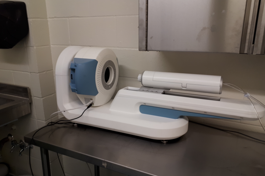
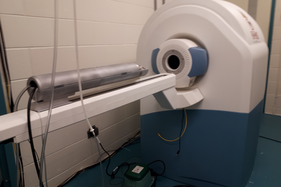
The core of the MRI component is a 7T cryogen free superconducting magnet with a 17 cm bore.
The cryogen free magnet imparts a small footprint and low mass to the MRI system, ensuring that it could be installed in our existing imaging facility with little or no renovation.
At the core of the PET component are two rings of dual layer silicon photomultiplier (lutetium-yttrium oxyorthosilicate) detectors forming a compact and robust PET imaging system that can be operated as a stand-alone PET imaging system or attached to the face of the MRI to operate as a sequential PET-MRI imaging system.
This new system therefore provides users the flexibility to conduct MR, PET, or PET-MR experiments.
Examples


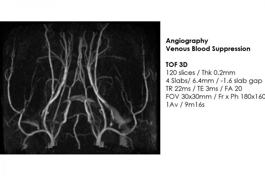
Instrumentation

Additional information
Any additional information? Please confirm.
Applications
Applications include:
Any additional information? Please confirm.
Resources
The materials listed here are available for investigators by request.
Adjusting RF calibration
Animal handling instructions
Image display user manual
MRS preclinical scan manual
PCSAM manual
PowerScan user manual
Reading noise level
Sequence library
Contact us
Small Animal and Material Imaging Core Facility (SAMICF)
23 Basic Medical Sciences Building
745 Bannatyne Avenue
University of Manitoba
Winnipeg, MB R3E 0J9 Canada