Overview
In conventional x-ray radiography, information is provided in only two dimensions, (x and y), Information relating to the third or depth (z) direction (corresponding to the direction of propagation of the incident x-rays) is lost as the three-dimensional object is collapsed into a two-dimensional image projected onto film or a detector. In CT imaging, this third dimension is preserved. This is achieved by obtaining a large number of observations at different viewing angles, which allows a cross sectional image to be produced by tomographic reconstruction.


Examples

Instrumentation
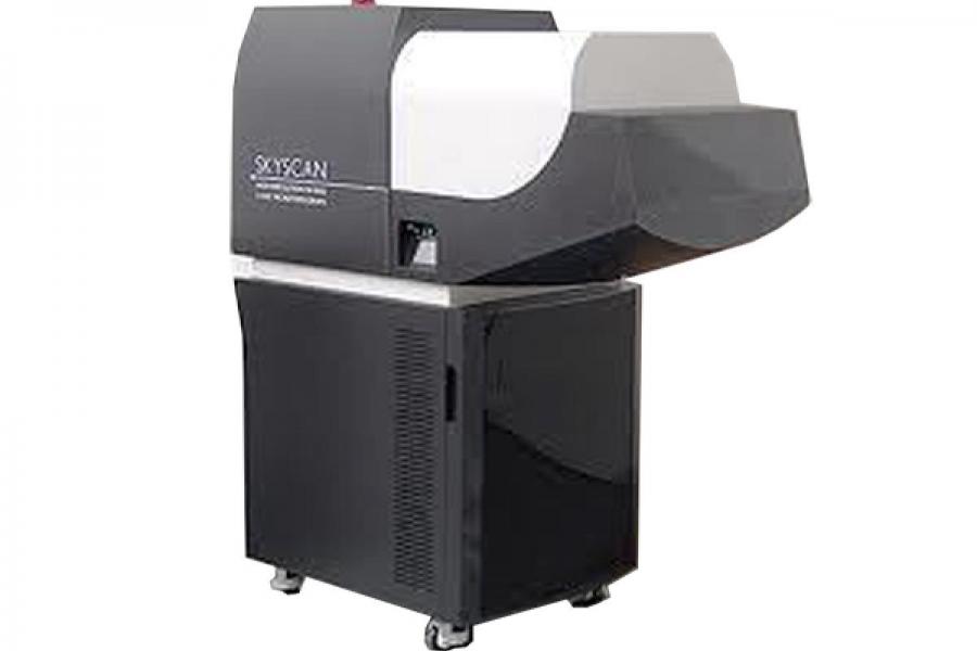
SkyScan 1176
CT images are acquired with a high performance SkyScan 1176 x-ray microtomography system equipped with a large format 11-megapixel x-ray camera.
Additional information
An image field width of up to 68 mm allows whole body scanning of rats and mice, with a scanning length of 20 to 200 mm possible.
Selectable image resolutions of 9, 18 and 35 um are available.
Variable x-ray applied voltage (20 to 90kV) and filters allow imaging of a wide range of samples in vivo from lung tissue to bone with titanium implants. The SkyScan 1176 is also well suited to study non-biological materials.
Image acquisition for a standard single field of view (22 mm by 38 mm) at 35 um resolution is seven minutes.
Typically, three fields of view are required to scan a whole mouse, giving a total acquisition time for a whole mouse scan at 35 um of 21 minutes.
Applications
Applications include:
- Non-invasive imaging of animal models of human disease
- Non-invasive imaging of genetically engineered animals
- Assess efficacy of novel pharmacological agents
- Assess novel drug delivery and gene therapy approaches
- Develop new contrast agents for diagnostic imaging
Resources
Application notes
The following Skyscan Application Notes are available to University of Manitoba staff and may be found on the data analysis workstations in BMSB 75 and 322:
3D visualization of open and closed porosity
3D volume analysis
Adaptive thresholding
Adipose tissue measurement in vivo
Adjacency CTAn 1
Adjacency CTAn 2
Advanced image co-registration in dataviewer
Advanced porosity analysis
Air bubbles in glass
Analysis of low-density material in a high-density container
Angular resampling
Anisotropy, MIL and stereology
Ant surface rendering for diamond
Automated trabecular and cortical bone selection
Basic 3D surface rendering
BMD calibration in CTAn
Bone micro-CT analysis general
Bone micro-CT analysis mouse
Bone micro-CT analysis rat
Bone sample scanning and analysis
Color coded 3D size distribution CTVol
Color coded 3D size distribution CTVox
Dual energy measurements
Embryo staining with PTA for ex vivo micro-CT imaging
Extracting open and closed pore networks for analysis and visualization
Heart ejection fraction analysis in vivo after synchronized scanning
How to set up a scan
Hu method note CTAn
Image and dataset registration in dataviewer
Image and dataset registration in dataviewer - expanded clay
Image and dataset registration in dataviewer - tooth
Image ROI for edited multi-part ROI
Impact of ROI on the analysis
Linear attenuation coefficients
Lung analysis in vivo after synchronized scanning
Minimal intensity projection in CTVox
MIP and minimal intensity projection of steel reinforced concrete
NRecon overview
Orientation analysis in 2D
Orthopedic micro-CT methods
Osteocyte and blood vessel analysis in cortical bone
Oversize reconstruction
Oversize reconstruction advanced
Porosity analysis
Rodent hind limb positioning for in vivo scan
Root canal treatment evaluation
Set binary selection as ROI
Setting pixel size
Sweep operation in 3D
Trajectory calibration
Where are left and right up and down in my images?
Bruker Micro-CT Academy
The materials listed here are available for investigators by request.
Bruker Micro-CT Academy 2014 Issue 1
Bruker Micro-CT Academy 2014 Issue 2
Bruker Micro-CT Academy 2014 Issue 3
Bruker Micro-CT Academy 2014 Issue 4
Bruker Micro-CT Academy 2014 Issue 5
Bruker Micro-CT Academy 2014 Issue 6
Bruker Micro-CT Academy 2014 Issue 7
Bruker Micro-CT Academy 2014 Issue 8
Bruker Micro-CT Academy 2014 Issue 9
Bruker Micro-CT Academy 2015 Issue 1
Bruker Micro-CT Academy 2015 Issue 2
Bruker Micro-CT Academy 2015 Issue 3
Bruker Micro-CT Academy 2015 Issue 4
Bruker Micro-CT Academy 2015 Issue 5
Bruker Micro-CT Academy 2015 Issue 6
Bruker Micro-CT Academy 2015 Issue 7
Bruker Micro-CT Academy 2015 Issue 8
Bruker Micro-CT Academy 2015 Issue 9
Bruker Micro-CT Academy 2014 Issue 10
Manuals
The materials listed here are available for investigators by request.
CTVol user manual
CTVox quick start guide
NRecon user manual
Skyscan 1176 user manual
Training videos
The following Skyscan training videos are available to University of Manitoba staff and may be found on the data analysis workstations in BMSB 75 and 322.
Adaptive thresholding of trabecular bone
BMD-TMD calibration for trabecular and cortical bone
Cortical bone 1 peri-endosteal volumes
Cortical bone 2 porosity
Cortical bone 3 peri-endosteal diameters
CTAn shrink-wrap
CTAn volume model viewing
New features CTAn 1-11
Setting the cortical VOI for mouse femur
Setting the trabecular VOI for mouse femur
Setting VOIs for rodent vertebra
Tooth part 1 pulp canal
Tooth part 2 enamel/dentine
Dataviewer training videos
Introduction to dataviewer
Re-orient a dataset in 3D with dataviewer
View cardiac gated scan in dataviewer
NRecon training videos
Adjusting xy ratio for oversize scan reconstruction
Resize projections in NRecon
YouTube videos
Larvae of paddlefish (Tetraodon) stained with phospho-tungstic acid
Maxilla and teeth of Saxon child, 1500 old archaeological specimen
Mouse maxilla and molar teeth, software auto-separation
Osteocyte lacunae and blood vessels in bone
Titanium implant in a pig rib, delineation of bone rings around implant surface
Contact us
Small Animal and Material Imaging Core Facility (SAMICF)
23 Basic Medical Sciences Building
745 Bannatyne Avenue
University of Manitoba
Winnipeg, MB R3E 0J9 Canada